Actinic Keratosis Risk Assessment Tool
This tool helps you understand your personal risk of developing Actinic Keratosis (AK) based on key risk factors. Select the factors that apply to you.
Your Risk Assessment
Select risk factors above to see your personalized risk assessment.
Important Information
People with multiple risk factors should consider annual skin examinations by a dermatologist. Early detection and treatment of Actinic Keratosis significantly reduces the risk of progression to skin cancer.
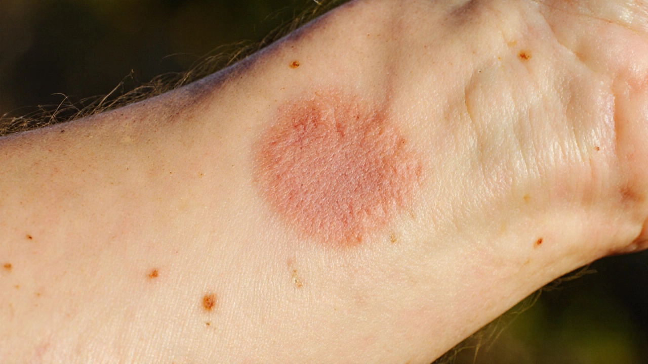
If you’ve noticed rough, scaly patches on sun‑exposed skin, you might be dealing with Actinic Keratosis. This condition sits at the crossroads of sun damage, changes in skin color, and a potential gateway to skin cancer. Below you’ll learn what it really is, why it messes with your skin’s pigment, and how to keep it under control.
Key Takeaways
- Actinic Keratosis (AK) is a sun‑induced precancerous growth that often appears as rough, pink‑ish spots.
- UV exposure triggers both AK and alterations in skin pigmentation, such as hyper‑ or hypopigmentation.
- Risk factors include age over 40, fair skin (Fitzpatrick I‑II), outdoor work, and a history of sunburns.
- Early diagnosis and treatment-cryotherapy, topical agents, or photodynamic therapy-greatly reduce the chance of progression to squamous cell carcinoma.
- Consistent sun protection (broad‑spectrum SPF30+, protective clothing, UV‑index awareness) prevents new lesions and limits pigment changes.
What Is Actinic Keratosis?
Actinic Keratosis is a rough, scaly growth that forms on skin chronically exposed to ultraviolet (UV) radiation. These lesions are typically pink, red, or flesh‑colored and feel sandpaper‑like to the touch. While most AKs stay harmless, about 1 in 100 can evolve into squamous cell carcinoma (SCC), a common form of skin cancer.
AK develops when UV‑induced DNA damage overwhelms the skin’s repair mechanisms. Over time, mutated cells accumulate in the outermost layer (the epidermis) and create the characteristic plaques.
How UV Exposure Changes Skin Pigmentation
Ultraviolet radiation covers UVA (320‑400nm) and UVB (280‑320nm) wavelengths that penetrate the skin and affect melanin production. Melanin, the pigment made by melanocytes, absorbs UV energy and protects deeper layers from damage. When UV exposure is excessive, two things happen:
- Hyperpigmentation: Melanocytes ramp up melanin synthesis, leading to dark patches (often called “sun spots” or “age spots”).
- Hypopigmentation: Repeated damage can destroy melanocytes, leaving lighter, sometimes whitish, areas.
Both outcomes are common in people with a history of AK, because the same UV insult that creates AK also triggers pigment shifts.
Link Between AK and Pigmentation Changes
When an AK is treated-especially with topical agents like 5‑fluorouracil (5‑FU) or imiquimod-the surrounding skin often reacts with inflammation. Once the inflammation fades, many patients notice post‑inflammatory hyperpigmentation (PIH). PIH appears as darker spots where the lesion was cleared and is more pronounced in skin types that naturally produce more melanin (Fitzpatrick III‑VI).
Conversely, cryotherapy (freezing the lesion with liquid nitrogen) can sometimes cause hypopigmented spots where melanocytes are damaged by the cold. Understanding these side effects helps set realistic expectations and guides after‑care, such as using gentle brightening moisturizers or sunscreen to minimize contrast.
Who Is Most at Risk?
Fitzpatrick skin type classifies skin based on its reaction to UV exposure is a quick way to gauge risk. Types I and II (very fair, often freckling, burns easily) develop AK earlier and more frequently. Other risk enhancers include:
- Age>40years-cellular repair slows down.
- Geography-living in sunny regions like Perth, Western Australia, where the UV index often exceeds 10.
- Outdoor occupations or hobbies (gardening, construction, surfing).
- History of blistering sunburns before age18.
- Immunosuppression (organ transplant recipients, chronic corticosteroid use).
If you tick several boxes, schedule a skin check at least once a year.
Diagnosis and When to See a Dermatologist
A dermatologist will start with a visual exam, often aided by a dermatoscope-a handheld magnifier that reveals vascular patterns unique to AK. When a lesion looks atypical, a shave or punch biopsy may be taken for histopathology.
Key signs that merit a professional visit:
- New or rapidly changing lesions on the face, scalp, ears, or hands.
- Any lesion that bleeds, crusts, or becomes painful.
- Existing AK that hasn’t responded to previous treatment.

Treatment Options: Choosing the Right Approach
Multiple therapies exist, each with pros and cons. Below is a quick comparison to help you pick what fits your skin, lifestyle, and budget.
| Treatment | How It Works | Typical Session | Success Rate | Common Side Effects |
|---|---|---|---|---|
| Cryotherapy | Liquid nitrogen freezes abnormal cells, causing them to slough off. | 1‑2minutes per lesion; often done in‑office. | 80‑90% clearance. | Redness, blistering, possible hypopigmentation. |
| Topical 5‑Fluorouracil (5‑FU) | Chemical agent that disrupts DNA synthesis in rapidly dividing cells. | Apply twice daily for 2‑4weeks. | 70‑85% clearance. | Severe inflammation, PIH, crusting. |
| Imiquimod Cream | Immune response modifier that boosts local interferon production. | Apply 5‑times per week for 6‑16weeks. | 60‑80% clearance. | Redness, itching, PIH. |
| Photodynamic Therapy (PDT) | Photosensitizing cream activated by a specific light wavelength destroys abnormal cells. | One‑hour clinic visit; may need repeat. | 85‑95% clearance. | Pain during illumination, erythema, temporary discoloration. |
For solitary lesions, cryotherapy is quick and cheap. For larger‑area AK (often on the scalp or forearms), topical creams or PDT provide a field‑treatment approach-treating both visible and sub‑clinical lesions.
Prevention: Sun‑Smart Habits That Matter
Sunscreen broad‑spectrum lotions that block UVA and UVB rays is the cornerstone of prevention. Choose SPF30 or higher, apply 15minutes before heading outdoors, and reapply every two hours-or after swimming or sweating.
Additional tactics:
- Wear UPF50+ clothing, wide‑brim hats, and UV‑blocking sunglasses.
- Check the daily UV index (available on weather apps). When it’s 7+, seek shade between 10a.m. and 4p.m.
- Use sunscreen on often‑overlooked spots: ears, neck, the back of the hands, and tops of the feet.
- Consider a vitaminD supplement if you limit sun exposure heavily.
Living With AK: Monitoring and Lifestyle Tips
Even after treatment, new AKs can appear. Adopt a “skin‑self‑check” routine:
- Stand in front of a well‑lit mirror.
- Inspect the face, scalp (use a handheld mirror), ears, neck, arms, and hands.
- Look for rough, scaly, or pigmented spots that differ from surrounding skin.
- Take a photo of any new lesion and note the date.
If something changes, book a dermatologist visit within a month. Also, keep a diary of sun exposure-knowing peak UV days helps you stay ahead of the curve.
Lastly, debunk common myths: AK is not contagious, it does not always turn into cancer, and over‑the‑counter acne creams are ineffective for these lesions.
Frequently Asked Questions
Can Actinic Keratosis go away on its own?
Rarely. Small AKs may flatten over months, but the underlying DNA damage remains. Without treatment, the risk of progression to squamous cell carcinoma persists.
Is it safe to use over‑the‑counter acne creams on AK?
No. Acne treatments target bacteria and oil, not the atypical cells in AK. Using them can irritate the skin further and mask the lesion, delaying proper care.
What’s the difference between hyperpigmentation after AK treatment and a new AK?
Post‑inflammatory hyperpigmentation (PIH) appears as a flat, darker spot where the skin healed. A new AK is usually raised or rough, sometimes pink, and feels like sandpaper. If you’re unsure, have a dermatologist examine it.
How often should I get a professional skin exam?
For individuals with a history of AK or high UV exposure, an annual full‑body skin exam is recommended. Those with multiple lesions may need check‑ups every 6months.
Can diet influence the development of AK?
While diet can’t replace sun protection, antioxidants (vitaminsC,E, selenium) help skin repair UV‑induced DNA damage. A balanced diet may modestly reduce risk.
Is photodynamic therapy painful?
Most patients describe a brief burning sensation during light activation, comparable to a mild sunburn. The discomfort usually fades within a few hours.
Bottom line: Actinic Keratosis is a warning sign that your skin has taken too much UV abuse. By spotting lesions early, treating them promptly, and committing to robust sun protection, you can keep both the lesions and unwanted pigment changes in check.
Comments (16)
Gerard Parker
Actinic keratosis (AK) is essentially a UV‑induced clonal proliferation of atypical keratinocytes that can serve as a premalignant field. The lesion’s rough, sand‑paper texture often betrays its presence long before any malignant transformation. UVB photons create cyclobutane pyrimidine dimers, while UVA generates oxidative stress; both pathways converge on DNA damage that overwhelms repair mechanisms. When the skin’s melanin shield is insufficient-typical of Fitzpatrick I‑II types-the damage accelerates, leading to hyper‑ or hypopigmented patches adjacent to AK. Epidemiological data consistently show that individuals over 40 with fair skin and outdoor occupations carry a three‑fold higher incidence. Immunosuppressed patients, such as organ‑transplant recipients, experience an even steeper risk curve because their surveillance pathways are blunted. Early detection hinges on a diligent self‑exam regimen: examine the face, scalp, ears, neck, dorsal hands, and forearms under good lighting every month. Dermatoscopic evaluation can differentiate the characteristic red‑purple vascular pattern of AK from benign actinic damage. When a lesion is confirmed, treatment options range from cryotherapy for isolated spots to field therapies like 5‑fluorouracil, imiquimod, or photodynamic therapy for multifocal disease. Cryotherapy boasts a rapid 80‑90 % clearance but may leave hypopigmented scars, especially in darker skin tones. Topical 5‑FU and imiquimod achieve comparable clearance rates but provoke intense inflammation that often results in post‑inflammatory hyperpigmentation (PIH). Photodynamic therapy offers the highest clearance (up to 95 %) yet requires specialized equipment and a brief painful illumination phase. Regardless of modality, adjunctive sunscreen use-broad‑spectrum SPF 30 or higher applied 15 minutes before exposure and reapplied every two hours-is non‑negotiable to prevent new lesions. Physical barriers such as UPF‑rated clothing, wide‑brim hats, and sunglasses further reduce cumulative UV dose. Nutritional antioxidants like vitamins C and E, as well as selenium, can modestly augment dermal repair but should never replace photoprotection. In summary, treat existing AK aggressively, protect irradiated skin relentlessly, and schedule annual dermatologist visits to stay ahead of the malignant edge.
Thomas Burke
Great rundown, thanks. I’ll definitely add a weekly sunscreen check to my routine.
Michelle Abbott
The pathophysiology you outlined is a textbook example of photodamage‑induced keratinocytic dysplasia.
Heather Jackson
OMG I totally felt that when I saw the same scaly plaques on my forearm after a beach trip, it was like my skin screaming for rescue!
Akshay Pure
While your summary captures the basics, it neglects the nuanced role of p53 mutations and the emerging data on field cancerization, which undoubtedly belong in any serious discourse on actinic keratosis.
Steven Macy
One could argue that AK serves as a reminder of our fragile equilibrium with nature; the sun offers life but also imposes a silent toll that we must respect.
Matt Stone
Skip the fancy creams just freeze it and move on
Joy Luca
Your description of PIH lacks precision; post‑inflammatory hyperpigmentation is a well‑characterised sequela of both 5‑FU and imiquimod therapy and should be addressed with topical hydroquinone or azelaic acid to mitigate contrast
Jessica Martins
The recommendation to reapply sunscreen every two hours is supported by pharmacokinetic studies showing a significant drop in SPF after 80 minutes of UV exposure.
Doug Farley
Oh sure, because everyone has a spare hour each day to stare at a mirror and catalog every speck of skin – that’s totally realistic.
Jeremy Olson
In accordance with current dermatological guidelines, individuals presenting with multiple actinic keratoses should be scheduled for biannual evaluations rather than the customary annual visit.
Ada Lusardi
I’ve finally learned that even a tiny dab of sunscreen can feel like a protective hug for my skin 🌞💖
Pam Mickelson
Keep up the diligent self‑checks; catching lesions early not only simplifies treatment but also dramatically lowers the risk of progression to squamous cell carcinoma.
Joe V
While your enthusiasm for sunscreen is commendable, let’s remember that SPF 50 does not make one invincible to a ten‑minute sunburn on a cloudless noon.
Scott Davis
Just remember to wear a hat.
Calvin Smith
If you think a single sunburn is scary, wait until you see the carnage a neglected AK can wreak on your dermis – it's the horror movie of dermatology, and the only plot twist is you actually had a chance to stop it.