Inflammation is a biological response to harmful stimuli that involves immune cells, blood vessels, and molecular mediators, characterized by redness, heat, swelling, and pain. It can be acute or chronic, with chronic inflammation persisting for months or years.
Osteoporosis is a skeletal disorder marked by reduced bone mineral density (BMD) and micro‑architectural deterioration, leading to heightened fracture risk. The condition silently progresses until a minor fall triggers a break.
Quick Take
- Chronic inflammation releases cytokines that boost bone‑resorbing cells.
- The RANKL‑OPG imbalance is the molecular bridge between the two conditions.
- Vitamin D, calcium, and anti‑inflammatory lifestyle choices can blunt the link.
- Targeted drugs such as denosumab and TNF‑α inhibitors show promise for bone preservation.
How Inflammation Works
When tissue injury or infection occurs, immune cells like macrophages and neutrophils converge on the site. They unleash a cocktail of signaling proteins-collectively called cytokines, including interleukins, tumor necrosis factor‑α (TNF‑α), and interferons-that coordinate the healing process.
In a healthy scenario, the inflammatory wave peaks quickly and then subsides, allowing tissue repair. In chronic disorders-rheumatoid arthritis, inflammatory bowel disease, or even persistent low‑grade metabolic inflammation-the cytokine surge stays elevated, keeping the immune system in overdrive.
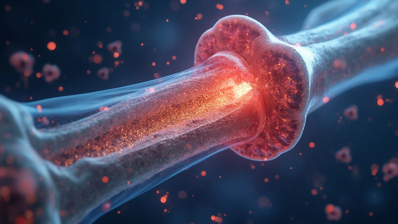
Bone Remodeling Basics
Bone isn’t static; it constantly remodels through a balance between two cell types:
- osteoclasts, the bone‑resorbing giants that break down old matrix.
- osteoblasts, the builders that lay down new collagen and mineral.
Under normal conditions, resorption and formation are tightly coupled, preserving bone strength. Disrupt that balance, and you tip toward loss (osteoporosis) or excess (osteopetrosis).
Cytokines That Bridge Inflammation and Bone Loss
Several cytokines have a direct hand in tipping the remodeling scale:
- TNF‑α accelerates osteoclast differentiation and suppresses osteoblast activity.
- Interleukin‑6 (IL‑6) enhances the expression of RANKL on stromal cells.
- Interleukin‑1 (IL‑1) triggers bone‑resorbing pathways similar to TNF‑α.
These molecules act like “inflam‑signals” that tell the skeletal system to break down more bone than it rebuilds, creating the inflammation osteoporosis link that clinicians now monitor.
The RANK/RANKL/OPG Pathway - The Molecular Bridge
The receptor activator of nuclear factor‑κB (RANK) sits on osteoclast precursors. Its ligand, RANKL, is produced by osteoblasts, stromal cells, and activated T‑cells during inflammation.
When RANKL binds RANK, it triggers a cascade that matures osteoclasts and prolongs their activity. The body’s natural brake, osteoprotegerin (OPG), acts as a decoy, soaking up excess RANKL.
Chronic inflammation raises RANKL while reducing OPG, skewing the balance toward bone loss. This shift is measurable in serum and serves as a predictive marker for fracture risk in patients with rheumatoid arthritis or systemic lupus.
| Attribute | Inflammation | Osteoporosis |
|---|---|---|
| Primary Cells | Macrophages, T‑cells | Osteoclasts > Osteoblasts |
| Key Mediators | TNF‑α, IL‑6, IL‑1 | RANKL ↑, OPG ↓ |
| Clinical Marker | CRP, ESR | BMD (g/cm²), FRAX score |
| Typical Treatment | NSAIDs, corticosteroids, biologics | Bisphosphonates, denosumab, calcium‑vit D |

Clinical Implications: Who’s at Risk?
Patients with long‑standing inflammatory diseases have a 1.5‑ to 2‑fold higher incidence of vertebral fractures compared to age‑matched controls. Post‑menopausal women with high‑sensitivity C‑reactive protein (hs‑CRP) above 3mg/L are especially vulnerable.
Beyond classic autoimmune conditions, metabolic inflammation linked to obesity and type‑2 diabetes also raises RANKL levels, making “silent” inflammation a hidden osteoporosis driver.
Nutrition and Lifestyle Strategies to Calm the Fire
Targeted diet and activity can lower inflammatory tone and protect bone:
- Omega‑3 fatty acids (e.g., salmon, walnuts) suppress TNF‑α production.
- Vitamin D (800-1000IU daily) enhances OPG expression and improves calcium absorption.
- Calcium (1000mg for adults) supplies the mineral scaffold.
- Weight‑bearing exercise (30min, 3times/week) stimulates osteoblasts and reduces visceral fat‑derived cytokines.
- Limit refined sugars and saturated fats, which trigger gut‑derived IL‑6.
The gut microbiome also talks to bone. Short‑chain fatty acids produced by fiber‑rich foods dampen systemic inflammation, indirectly supporting bone density.
Emerging Therapies Targeting Inflammatory Bone Loss
Pharmacology is moving beyond generic anti‑resorptives:
- Denosumab is a monoclonal antibody that mimics OPG, binding RANKL and curbing osteoclast activity. Clinical trials in rheumatoid arthritis showed a 40% reduction in vertebral fractures.
- Biologics that neutralize TNF‑α (e.g., etanercept, infliximab) not only ease joint pain but also improve BMD scores over a 2‑year horizon.
- Selective cytokine inhibitors targeting IL‑6 (tocilizumab) have demonstrated modest gains in hip BMD in osteopenic patients.
Future directions include small‑molecule RANKL antagonists and gene‑editing approaches that up‑regulate OPG production.
Related Concepts
Understanding the inflammation‑osteoporosis connection also sheds light on other health arenas:
- Cardiovascular disease shares the same cytokine milieu; high IL‑6 predicts both atherosclerosis and bone loss.
- Chronic kidney disease‑mineral bone disorder (CKD‑MBD) illustrates how systemic inflammation can derail calcium‑phosphate metabolism.
- Periodontal disease serves as a localized model where bacterial inflammation accelerates alveolar bone resorption via RANKL.
Exploring these links helps clinicians adopt a holistic view: treat the inflammation, and the bone often follows.
Frequently Asked Questions
Can short‑term inflammation cause osteoporosis?
Acute inflammation alone usually doesn’t lead to measurable bone loss. It’s the prolonged, low‑grade inflammation that continuously elevates RANKL and depresses OPG, gradually eroding bone density over months to years.
Do NSAIDs protect my bones?
NSAIDs blunt prostaglandin‑mediated inflammation, which can modestly reduce osteoclast activation. However, chronic high‑dose use may impair kidney function and calcium balance, so they’re not a long‑term bone‑saving strategy.
Is the RANKL test available for patients?
Serum RANKL assays exist but are mainly used in research settings. Clinicians more commonly rely on indirect markers like CRP, ESR, and DXA‑derived BMD scores to gauge fracture risk.
How much vitamin D do I need to counter inflammation?
Most guidelines suggest 800-1000IU daily for adults, aiming for serum 25‑OH vitamin D levels of 30-50ng/mL. This range supports OPG production and dampens cytokine spikes.
Can weight‑bearing exercise reverse bone loss caused by inflammation?
Regular weight‑bearing activity stimulates osteoblasts and can increase BMD by 1‑3% over a year, even in inflammatory conditions. Pairing exercise with anti‑inflammatory nutrition yields the best results.
Comments (13)
Kelvin Murigi
Great synthesis of the inflammation‑osteoporosis axis! You nailed the RANKL‑OPG imbalance and highlighted how chronic cytokine exposure skews bone remodeling. I’d add that even low‑grade adipose‑derived IL‑6 can tip the scale in sedentary folks. Also, ensuring adequate vitamin D isn’t just about calcium – it up‑regulates OPG, closing that feedback loop you described. Keep the clinical pearls coming; they’re gold for primary‑care docs.
ahmad matt
Honestly this is just fluff.
kristine ayroso
Okay, let me break this down because there are a ton of moving parts here. First off, chronic inflammation isn’t just a side effect of some autoimmune disease – it’s a whole ecosystem of immune signaling that hijacks bone homeostasis. When you have persistent TNF‑α, IL‑1, and IL‑6 around, you’re basically telling osteoclast precursors to grow up faster and act like they own the place. At the same time, those same cytokines put the brakes on osteoblasts, so you get a double whammy of bone loss. The RANKL‑OPG balance is the key lever – RANKL goes up, OPG goes down, and you end up with a net increase in bone resorption. It’s not just theory; you can measure elevated serum RANKL in rheumatoid arthritis patients and see it correlate with fracture risk. Then there’s the metabolic angle: obesity fuels low‑grade inflammation via adipokines, and diabetes throws advanced glycation end‑products into the mix, further raising RANKL expression. Nutrition matters too – omega‑3s knock down TNF‑α production, while vitamin D pushes OPG up. And don’t forget exercise; mechanical loading stimulates osteoblast activity and can blunt the cytokine surge coming from visceral fat. On the therapeutic front, denosumab literally mimics OPG and has shown a 40 % drop in vertebral fractures in RA trials, which is huge. TNF inhibitors do double duty by easing joint pain and nudging BMD up over a couple of years. IL‑6 blockers are still catching up, but early data looks promising for hip density. All that said, we need to think holistically – treat the inflammation, adjust diet, get patients moving, and consider bone‑specific drugs when the risk is high. That’s the only way to keep the skeleton from crumbling under a chronic fire.
Ben Small
Spot on. The interplay is vicious, and without aggressive management you’ll lose bone faster than you think.
Dylan Hilton
Exactly, and to add, many patients don’t realize a simple tweak like adding a three‑serving fish schedule can shave off months of bone loss. Pair that with a daily vitamin D dose and you create a protective shield around the RANKL‑OPG axis.
Christian Andrabado
Interesting points. However, be careful with high‑dose NSAIDs; they may harm kidney function and calcium handling.
Chidi Anslem
True, moderation is key. Balancing anti‑inflammatory meds with lifestyle changes yields the best outcomes without over‑relying on pharmaceuticals.
Holly Hayes
Nice summary but you totally missed how glucocorticoids slam the OPG production.
Penn Shade
This post completely ignores the role of glucocorticoids.
Jennifer Banash
While the article is comprehensive, I would caution readers to consult their physicians before initiating any supplementation regimen, as excessive calcium can precipitate vascular calcifications in susceptible individuals.
Stephen Gachie
One could argue that focusing solely on cytokines oversimplifies the complex endocrine feedback loops involving parathyroid hormone and fibroblast growth factor‑23, which also modulate bone turnover under inflammatory conditions.
Sara Spitzer
Good read, but the data on omega‑3s is still mixed; some trials show no significant BMD benefit, so don’t hype it too much.
Emily Wang
Bottom line: chronic inflammation is a silent bone thief – tackle it with meds, diet, and exercise, or risk fractures.